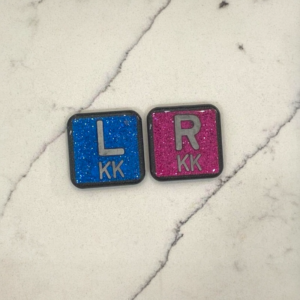
If you have ever had an X-ray taken, you may have noticed a small letter tucked into the corner of the image, either an R or an L. It is easy to overlook, but that tiny marker is doing a lot of work. As a Registered X-ray Technologist with over 20 years of experience, I place these markers on every single patient I image, and I want to walk you through exactly why.
The Marker Is Part of the Tool Kit We Bring to Work Every Day
Radiographers do not just show up with a machine. We show up with our markers. These are small pieces, usually about an inch in size, encased in durable resin so they can be cleaned between patients. Every technologist has a set with their own initials built in. We place one beside the body part before or during the exposure so the image clearly shows which side of your body was imaged, your right or your left.
Why This Matters Legally
An X-ray is not just a picture. It is a legal document. That means the R or L marker has to be visible on the image, along with the radiographer’s initials. This creates a permanent record of exactly who imaged you, which side of your body was examined, and how the image was taken. If there is ever a question down the road about your care, that marker and those initials are part of the proof.
Why This Matters for Your Diagnosis
Beyond the legal side, the marker plays a real role in how your doctor and the radiologist interpret your images.
Take one of the most common X-rays we perform, a PA chest and lateral chest X-ray. If you are able to stand, the image is taken with the L marker placed on the imaging plate while you face the machine. Your heart normally sits on the left side of your chest.
Here is where it gets interesting. If the marker was placed correctly and the heart appears on the right side of the image instead of the left, that is not a marker error. That could be a sign of dextrocardia or situs inversus, a rare congenital condition where a person’s internal organs are a complete mirror image of typical anatomy. The heart points to the right, and organs like the liver, stomach, and spleen are all switched to the opposite side of where they are usually found.
What makes this especially compelling is that some patients do not even know they have this condition until an X-ray reveals it. That tiny marker can be the first clue in uncovering something significant about a person’s own body.
One Small Detail That Changes Everything
The orientation of the marker matters just as much as the letter itself. If a patient is positioned prone, lying face down, the right and left sides are reversed on the imaging plate compared to when someone is standing or lying face up. Technologists are trained to account for this before the image is even taken, not after. This is one of those details that separates a properly trained radiographer from someone just pointing a machine at a body part.
What Happens When a Marker Is Wrong
Mistakes can happen. If an X-ray of a right knee accidentally gets an L marker placed on it, anatomy itself will often tell the true story. The fibula, one of the two lower leg bones, always sits on the lateral, or outer, side of the leg because it is not a weight bearing bone. A radiologist or radiographer looking at that image can usually recognize right away that the marker does not match the anatomy.
This is exactly why so much time in medical radiography school, and in the clinical environment afterward, is spent drilling marker placement. It is not a small detail to us. It is treated as a core skill.
Markers Are Used Beyond Standard X-Ray
Markers are not just an X-ray tool. Other imaging teams use them too. Occupational therapists performing swallow studies under fluoroscopy use markers labeled thin, thick, and solid. As the patient swallows different textures during the study, the OT can watch in real time and see exactly which textures the patient can safely tolerate. It is a great example of how a simple physical marker can guide a much more complex clinical decision.
A Small Story From the Business Side
Through XrayMarkers.ca, I get to hear directly from technologists about how they use their markers day to day. One request has always stuck with me. A radiographer asked me to make her markers as plain as possible, no fun colors or designs, because a previous patient had liked her markers so much that they took one home as a souvenir. It is a funny reminder that even the smallest tools we use every day can leave an impression.
What I Want You to Take Away
The next time you have an X-ray taken and you notice that small R or L marker in the corner of the image, I hope you see it differently. It is not decoration. It is a legal safeguard, a diagnostic clue, and a tool that every radiographer carries with them for every single patient, every single day.